Genetics - MCAT Biology
Card 0 of 20
A diploid human cell that is dividing will contain _______ chromosomes. These chromosomes will each consist of _______ chromatids. Fill in the corresponding blanks.
A diploid human cell that is dividing will contain _______ chromosomes. These chromosomes will each consist of _______ chromatids. Fill in the corresponding blanks.
The diploid number is 46 and the haploid number is 23. When cells are dividing, each chromosome is present in duplicate copy. These chromosomes are composed of two chromatids each when they are replicated.
The diploid number is 46 and the haploid number is 23. When cells are dividing, each chromosome is present in duplicate copy. These chromosomes are composed of two chromatids each when they are replicated.
Compare your answer with the correct one above
Human chromosomes are divided into two arms, a long q arm and a short p arm. A karyotype is the organization of a human cell’s total genetic complement. A typical karyotype is generated by ordering chromosome 1 to chromosome 23 in order of decreasing size.
When viewing a karyotype, it can often become apparent that changes in chromosome number, arrangement, or structure are present. Among the most common genetic changes are Robertsonian translocations, involving transposition of chromosomal material between long arms of certain chromosomes to form one derivative chromosome. Chromosomes 14 and 21, for example, often undergo a Robertsonian translocation, as below.

A karyotype of this individual for chromosomes 14 and 21 would thus appear as follows:

Though an individual with aberrations such as a Robertsonian translocation may be phenotypically normal, they can generate gametes through meiosis that have atypical organizations of chromosomes, resulting in recurrent fetal abnormalities or miscarriages.
In der(14,21), which region of a chromosome might you expect to find in the center of its structure?
Human chromosomes are divided into two arms, a long q arm and a short p arm. A karyotype is the organization of a human cell’s total genetic complement. A typical karyotype is generated by ordering chromosome 1 to chromosome 23 in order of decreasing size.
When viewing a karyotype, it can often become apparent that changes in chromosome number, arrangement, or structure are present. Among the most common genetic changes are Robertsonian translocations, involving transposition of chromosomal material between long arms of certain chromosomes to form one derivative chromosome. Chromosomes 14 and 21, for example, often undergo a Robertsonian translocation, as below.
A karyotype of this individual for chromosomes 14 and 21 would thus appear as follows:
Though an individual with aberrations such as a Robertsonian translocation may be phenotypically normal, they can generate gametes through meiosis that have atypical organizations of chromosomes, resulting in recurrent fetal abnormalities or miscarriages.
In der(14,21), which region of a chromosome might you expect to find in the center of its structure?
Telomeres are present at the ends of chromosomes. If one really understands the passage, one can see that Robertsonian translocation places the ends of chromosomes together to form the middle of the derivative chromosome. We would expect to find telomeres in this region.
Telomeres are present at the ends of chromosomes. If one really understands the passage, one can see that Robertsonian translocation places the ends of chromosomes together to form the middle of the derivative chromosome. We would expect to find telomeres in this region.
Compare your answer with the correct one above
Human chromosomes are divided into two arms, a long q arm and a short p arm. A karyotype is the organization of a human cell’s total genetic complement. A typical karyotype is generated by ordering chromosome 1 to chromosome 23 in order of decreasing size.
When viewing a karyotype, it can often become apparent that changes in chromosome number, arrangement, or structure are present. Among the most common genetic changes are Robertsonian translocations, involving transposition of chromosomal material between long arms of certain chromosomes to form one derivative chromosome. Chromosomes 14 and 21, for example, often undergo a Robertsonian translocation, as below.
A karyotype of this individual for chromosomes 14 and 21 would thus appear as follows:
Though an individual with aberrations such as a Robertsonian translocation may be phenotypically normal, they can generate gametes through meiosis that have atypical organizations of chromosomes, resulting in recurrent fetal abnormalities or miscarriages.
In a normal chromosome 14, what region of the chromosome exists between the p arm and the q arm?
In a normal chromosome, the passage indicates that the p and q arm meet in the center. This central region of the chromosome is known as a centromere.
In a normal chromosome, the passage indicates that the p and q arm meet in the center. This central region of the chromosome is known as a centromere.
Compare your answer with the correct one above
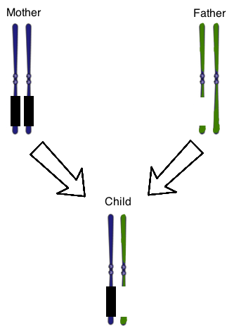
The concept of genomic imprinting is important in human genetics. In genomic imprinting, a certain region of DNA is only expressed by one of the two chromosomes that make up a typical homologous pair. In healthy individuals, genomic imprinting results in the silencing of genes in a certain section of the maternal chromosome 15. The DNA in this part of the chromosome is "turned off" by the addition of methyl groups to the DNA molecule. Healthy people will thus only have expression of this section of chromosome 15 from paternally-derived DNA.
The two classic human diseases that illustrate defects in genomic imprinting are Prader-Willi and Angelman Syndromes. In Prader-Willi Syndrome, the section of paternal chromosome 15 that is usually expressed is disrupted, such as by a chromosomal deletion. In Angelman Syndrome, maternal genes in this section are deleted, while paternal genes are silenced. Prader-Willi Syndrome is thus closely linked to paternal inheritance, while Angelman Syndrome is linked to maternal inheritance.
Figure 1 shows the chromosome 15 homologous pair for a child with Prader-Willi Syndrome. The parental chromosomes are also shown. The genes on the mother’s chromosomes are silenced normally, as represented by the black boxes. At once, there is also a chromosomal deletion on one of the paternal chromosomes. The result is that the child does not have any genes expressed that are normally found on that region of this chromosome.
Chromosome 15 is an autosome. Which of the following is (are) true of all autosomes?
I. They contain histones
II. They determine chromosomal sex
III. They align on the metaphase plate during mitosis
Autosomes are the chromosomes that are not sex chromosomes. Any numbered chromosome (1 through 22) is an autosome, while the X and Y chromosomes (the 23rd pair) are the sex chromosomes. Statement II is only true of the X and Y chromosomes. Statements I and III are true of all chromosomes.
Autosomes are the chromosomes that are not sex chromosomes. Any numbered chromosome (1 through 22) is an autosome, while the X and Y chromosomes (the 23rd pair) are the sex chromosomes. Statement II is only true of the X and Y chromosomes. Statements I and III are true of all chromosomes.
Compare your answer with the correct one above
The concept of genomic imprinting is important in human genetics. In genomic imprinting, a certain region of DNA is only expressed by one of the two chromosomes that make up a typical homologous pair. In healthy individuals, genomic imprinting results in the silencing of genes in a certain section of the maternal chromosome 15. The DNA in this part of the chromosome is "turned off" by the addition of methyl groups to the DNA molecule. Healthy people will thus only have expression of this section of chromosome 15 from paternally-derived DNA.
The two classic human diseases that illustrate defects in genomic imprinting are Prader-Willi and Angelman Syndromes. In Prader-Willi Syndrome, the section of paternal chromosome 15 that is usually expressed is disrupted, such as by a chromosomal deletion. In Angelman Syndrome, maternal genes in this section are deleted, while paternal genes are silenced. Prader-Willi Syndrome is thus closely linked to paternal inheritance, while Angelman Syndrome is linked to maternal inheritance.
Figure 1 shows the chromosome 15 homologous pair for a child with Prader-Willi Syndrome. The parental chromosomes are also shown. The genes on the mother’s chromosomes are silenced normally, as represented by the black boxes. At once, there is also a chromosomal deletion on one of the paternal chromosomes. The result is that the child does not have any genes expressed that are normally found on that region of this chromosome.
Imagine the child in figure 1 was diagnosed at birth with cystic fibrosis as well as Prader-Willi. Cystic fibrosis is due to a recessive genetic mutation on chromosome 7. Two years later, his parents have another child that has cystic fibrosis, but not Prader-Willi. Which of the following best explains why Prader-Willi and cystic fibrosis are not always inherited together ?
The law of independent assortment says that chromosomes, and thus most genes, align independently of each other when being passed from parent to child. In other words, chromosome 7 and chromosome 15 do not directly influence each other's inheritance patterns during meiosis in parental gametes, and can be sent to sperm or eggs in any combination.
The law of independent assortment says that chromosomes, and thus most genes, align independently of each other when being passed from parent to child. In other words, chromosome 7 and chromosome 15 do not directly influence each other's inheritance patterns during meiosis in parental gametes, and can be sent to sperm or eggs in any combination.
Compare your answer with the correct one above
The concept of genomic imprinting is important in human genetics. In genomic imprinting, a certain region of DNA is only expressed by one of the two chromosomes that make up a typical homologous pair. In healthy individuals, genomic imprinting results in the silencing of genes in a certain section of the maternal chromosome 15. The DNA in this part of the chromosome is "turned off" by the addition of methyl groups to the DNA molecule. Healthy people will thus only have expression of this section of chromosome 15 from paternally-derived DNA.
The two classic human diseases that illustrate defects in genomic imprinting are Prader-Willi and Angelman Syndromes. In Prader-Willi Syndrome, the section of paternal chromosome 15 that is usually expressed is disrupted, such as by a chromosomal deletion. In Angelman Syndrome, maternal genes in this section are deleted, while paternal genes are silenced. Prader-Willi Syndrome is thus closely linked to paternal inheritance, while Angelman Syndrome is linked to maternal inheritance.
Figure 1 shows the chromosome 15 homologous pair for a child with Prader-Willi Syndrome. The parental chromosomes are also shown. The genes on the mother’s chromosomes are silenced normally, as represented by the black boxes. At once, there is also a chromosomal deletion on one of the paternal chromosomes. The result is that the child does not have any genes expressed that are normally found on that region of this chromosome.
Based on the information in the passage, which of the following is true of Prader-Willi Syndrome?
I. It must involve a chromosomal deletion on the paternal chromosome 15
II. It must involve normal silencing of maternal chromosome 15
III. It is a sex-linked disorder because it involves chromosome 15
Prader-Willi must involve the silencing of maternal genes on chromosome 15, as well as some disruption of the paternal chromosome. A chromosomal deletion is one example of this kind of disruption, but could also be a nonsense mutation or frameshift mutation that renders the paternal DNA unable to be ultimately translated to protein.
Sex-linked disorders involve the X and Y chromosomes. Prader-Willi is inherited through chromosome 15, and is thus not sex-linked.
Compare your answer with the correct one above
A man with type AB blood marries a woman with type A blood. Which of the following blood types might their sons inherit?
A man with type AB blood marries a woman with type A blood. Which of the following blood types might their sons inherit?
The genotype of the father is definitely AB. The genotype of the mother is either AA or AO.
Below are the Punnett squares that show that types A, B, and AB are possible. Note that the genotype AO will result in a type A phenotype, while the genotype BO will result in a type B phenotype.

The genotype of the father is definitely AB. The genotype of the mother is either AA or AO.
Below are the Punnett squares that show that types A, B, and AB are possible. Note that the genotype AO will result in a type A phenotype, while the genotype BO will result in a type B phenotype.
Compare your answer with the correct one above
Chromosomal aberrations, such as trisomy or monosomy, are often the result of nondisjunction during cell division. Nondisjunction is characterized by a malfunction during which stage of division?
Chromosomal aberrations, such as trisomy or monosomy, are often the result of nondisjunction during cell division. Nondisjunction is characterized by a malfunction during which stage of division?
This question is asking about the phase of Meiosis, during which disjunction (separation) of chromosomes normally occurs. While Metaphase can sometimes be a seductive answer, note that disjunction officially occurs during Anaphase.
This question is asking about the phase of Meiosis, during which disjunction (separation) of chromosomes normally occurs. While Metaphase can sometimes be a seductive answer, note that disjunction officially occurs during Anaphase.
Compare your answer with the correct one above
Which answer choice correctly shows a frameshift mutation?
Which answer choice correctly shows a frameshift mutation?
A framshift mutation is when an extra base is inserted into (or deleted from) a strand of DNA. When the DNA is transcribed, every codon downstream from the insertion is changed because the reading frame will be different. This will be the case for any mutations that affect a sequence of nucleotides that is not a multiple of three, for example a five-nucleotide deletion. Such mutations affect the three-nucleotide groupings for all codons following the mutation, and often result in a premature stop codon.
The correct answer is CGGTGAATAGGC  CGGTGAATCAGGC. In the original sequence, the codons are CGG-TGA-ATA-GGC. In the second, they become CGG-TGA-ATC-AGG-C. The other answers show either insertions or deletions that do not shift the reading frame, or single nucleotide polymorphism (changing a single nucleotide to another).
Note that the reading frame does not change in the following 3-nucleotide insertion. Though there is an extra codon, the downstream sequence is not affected.
CGG-TGA-ATA-GGC  CGG-TGA-AGA-CTA-GGC
A framshift mutation is when an extra base is inserted into (or deleted from) a strand of DNA. When the DNA is transcribed, every codon downstream from the insertion is changed because the reading frame will be different. This will be the case for any mutations that affect a sequence of nucleotides that is not a multiple of three, for example a five-nucleotide deletion. Such mutations affect the three-nucleotide groupings for all codons following the mutation, and often result in a premature stop codon.
The correct answer is CGGTGAATAGGC
Note that the reading frame does not change in the following 3-nucleotide insertion. Though there is an extra codon, the downstream sequence is not affected.
CGG-TGA-ATA-GGC
Compare your answer with the correct one above
Human chromosomes are divided into two arms, a long q arm and a short p arm. A karyotype is the organization of a human cell’s total genetic complement. A typical karyotype is generated by ordering chromosome 1 to chromosome 23 in order of decreasing size.
When viewing a karyotype, it can often become apparent that changes in chromosome number, arrangement, or structure are present. Among the most common genetic changes are Robertsonian translocations, involving transposition of chromosomal material between long arms of certain chromosomes to form one derivative chromosome. Chromosomes 14 and 21, for example, often undergo a Robertsonian translocation, as below.
A karyotype of this individual for chromosomes 14 and 21 would thus appear as follows:
Though an individual with aberrations such as a Robertsonian translocation may be phenotypically normal, they can generate gametes through meiosis that have atypical organizations of chromosomes, resulting in recurrent fetal abnormalities or miscarriages.
During meiosis, gametes result from the isolation of one chromosome from each of a human’s homologous 23 pairs. In Figure 2, der(14,21) is treated as a homologous pair to either 14 or 21. An egg, then, could be formed that had a normal chromosome 21 and the der(14,21) instead of the normal chromosome 14. If a sperm fertilizes this egg, and the sperm has a normal chromosome 14 and 21, what will result?
Since the maternal oocyte would have two copies of chromosome 21, the normal 21 and the derivative chromosome, the normal addition of the sperm would lead to a zygote with a total of three copies of chromosome 21, or trisomy 21. Polyploidy is a tempting choice, but polyploid is a state characterized by extra copies of multiple chromosomes, as might be expected from more than one sperm fertilizing an egg.
Since the maternal oocyte would have two copies of chromosome 21, the normal 21 and the derivative chromosome, the normal addition of the sperm would lead to a zygote with a total of three copies of chromosome 21, or trisomy 21. Polyploidy is a tempting choice, but polyploid is a state characterized by extra copies of multiple chromosomes, as might be expected from more than one sperm fertilizing an egg.
Compare your answer with the correct one above
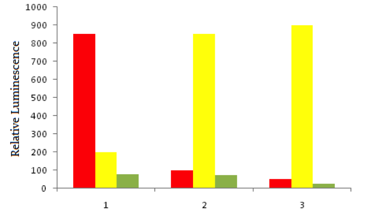
Scientists use a process called Flourescent In-Situ Hybridization, or FISH, to study genetic disorders in humans. FISH is a technique that uses spectrographic analysis to determine the presence or absence, as well as the relative abundance, of genetic material in human cells.
To use FISH, scientists apply fluorescently-labeled bits of DNA of a known color, called probes, to samples of test DNA. These probes anneal to the sample DNA, and scientists can read the colors that result using laboratory equipment. One common use of FISH is to determine the presence of extra DNA in conditions of aneuploidy, a state in which a human cell has an abnormal number of chromosomes. Chromosomes are collections of DNA, the totality of which makes up a cell’s genome. Another typical use is in the study of cancer cells, where scientists use FISH labels to ascertain if genes have moved inappropriately in a cell’s genome.
Using red fluorescent tags, scientists label probe DNA for a gene known to be expressed more heavily in cancer cells than normal cells. They then label a probe for an immediately adjacent DNA sequence with a green fluorescent tag. Both probes are then added to three dishes, shown below. In dish 1 human bladder cells are incubated with the probes, in dish 2 human epithelial cells are incubated, and in dish 3 known non-cancerous cells are used. The relative luminescence observed in regions of interest in all dishes is shown below.
Which of the following genetic changes would not be detectable using FISH?
FISH is useful for visualizing major changes in the genome, as described by the passage. Translocations and large copy number variants would likley be visible in color changes, while small single nucleotide polymorphisms (SNPs) would likely be less easily detected.
FISH is useful for visualizing major changes in the genome, as described by the passage. Translocations and large copy number variants would likley be visible in color changes, while small single nucleotide polymorphisms (SNPs) would likely be less easily detected.
Compare your answer with the correct one above
Cryptosporidium is a genus of gastrointestinal parasite that infects the intestinal epithelium of mammals. Cryptosporidium is water-borne, and is an apicomplexan parasite. This phylum also includes Plasmodium, Babesia, and Toxoplasma.
Apicomplexans are unique due to their apicoplast, an apical organelle that helps penetrate mammalian epithelium. In the case of cryptosporidium, there is an interaction between the surface proteins of mammalian epithelial tissue and those of the apical portion of the cryptosporidium infective stage, or oocyst. A scientist is conducting an experiment to test the hypothesis that the oocyst secretes a peptide compound that neutralizes intestinal defense cells. These defense cells are resident in the intestinal epithelium, and defend the tissue by phagocytizing the oocysts.
She sets up the following experiment:
As the neutralizing compound was believed to be secreted by the oocyst, the scientist collected oocysts onto growth media. The oocysts were grown among intestinal epithelial cells, and then the media was collected. The media was then added to another plate where Toxoplasma gondii was growing with intestinal epithelial cells. A second plate of Toxoplasma gondii was grown with the same type of intestinal epithelium, but no oocyst-sourced media was added.
You are conducting a study of an isolated tribe in New Guinea, and you find that there is widespread resistance to cryptosporidium infection. Upon examination, you find that the resistance is caused by a change in one nucleotide pair in a gene on chromosome 13. What kind of genetic change does this likely reflect?
This would be an example of a single nucleotide polymorphism. A fairly common variant is some change at a single base pair in human DNA. It is possible that this base pair change results in a modified protein that functions just as well as the normal protein in most conditions.
Some of these changes may, simply by chance, be better at resisting disease. When exposed to stress, such as a disease epidemic, it is possible this variant of normal becomes the most widespread genotype. This is especially noticeable in an isolated population, like a tribe in New Guinea. Note that not all single nucleotide polymorphisms will have positive effects, and in some cases can cause disease instead of resistance.
Compare your answer with the correct one above
An mRNA sequence is supposed to read UAUGGA, but a mutation replaces the second uracil base with guanine. What is the most specific term for this type of mutation?
An mRNA sequence is supposed to read UAUGGA, but a mutation replaces the second uracil base with guanine. What is the most specific term for this type of mutation?
This mutation replaced UAU (a coding codon) with UAG (a stop codon). The most descriptive term for this kind of replacement is a nonsense mutation.
Note that an easy way to remember the stop codons is UGA ("u" get away), UAA ("u" are away), and UAG ("u" are gone).
This mutation replaced UAU (a coding codon) with UAG (a stop codon). The most descriptive term for this kind of replacement is a nonsense mutation.
Note that an easy way to remember the stop codons is UGA ("u" get away), UAA ("u" are away), and UAG ("u" are gone).
Compare your answer with the correct one above
A mutation within a gene results in the premature addition of a stop codon during translation. This describes which type of mutation?
A mutation within a gene results in the premature addition of a stop codon during translation. This describes which type of mutation?
A nonsense mutation results in premature addition of a stop codon. A missense mutation is a point mutation that results in a different codon, which ultimately codes for a different amino acid in the polypetide sequence. A frameshift mutation results from and insertion or deletion of a nucleotide, resulting in a change in the reading frame. Frameshift mutations often indirectly result in nonsense mutations, but are not the best answer choice given.
A nonsense mutation results in premature addition of a stop codon. A missense mutation is a point mutation that results in a different codon, which ultimately codes for a different amino acid in the polypetide sequence. A frameshift mutation results from and insertion or deletion of a nucleotide, resulting in a change in the reading frame. Frameshift mutations often indirectly result in nonsense mutations, but are not the best answer choice given.
Compare your answer with the correct one above
Imagine there is a mutation in a gene where a nucleotide was replaced by another nucleotide. The mutation did not affect the primary structure of the protein for which the gene coded.
What type of mutation is this?
Imagine there is a mutation in a gene where a nucleotide was replaced by another nucleotide. The mutation did not affect the primary structure of the protein for which the gene coded.
What type of mutation is this?
Whenever a base pair is replaced by another base pair in the DNA double helix, it is referred to as a point mutation. If the amino acid is not changed by the mutation, we can refer to the mutation as a silent mutation, as the primary protein structure is not affected.
Point mutation: CATGA becomes CAGGA
Insertion mutations occur when additional bases are inserted into the DNA sequence.
Insertion mutation: CATGA becomes CATACTGA
Frameshift mutations can result from insertions or deletions when the number of nucleotides added/removed is not a multiple of three. Since codons are grouped by threes, any change that is not a multiple of three will alter the grouping of every codon downstream of the mutation, severly altering the primary protein structure.
Frameshift mutation: CATGA becomes CAATGA
A nonsense mutation results in a premature stop codon, and early translation termination. This can arise from a frameshift mutation, point mutation, insertion, or deletion.
Nonsense mutation: CATGA becomesCATTTAGA (When transcribed, this sequence becomes the mRNA UCUAAAUG, where UAA is a stop codon).
Compare your answer with the correct one above
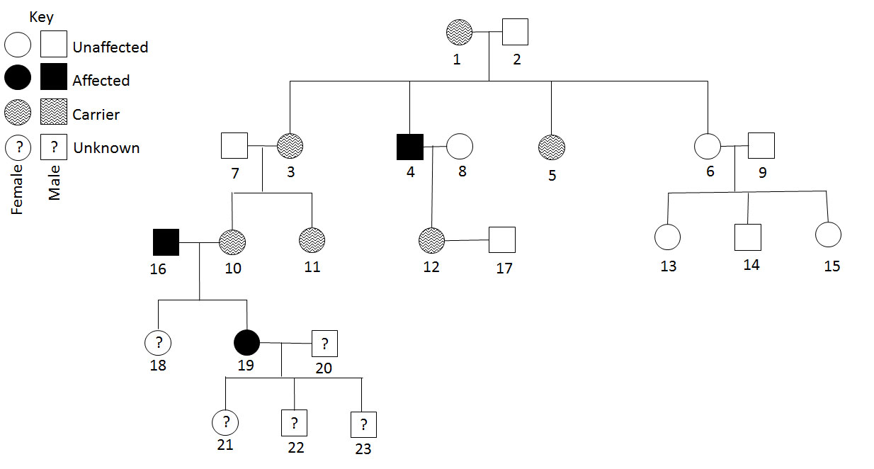
Consider the pedigree. Is the trait dominant or recessive?
Consider the pedigree. Is the trait dominant or recessive?
The trait is recessive because affected individuals do not occur in every generation. Additionally, dominant traits do not result in "carriers". Individuals are either affected or unaffected.
The trait is recessive because affected individuals do not occur in every generation. Additionally, dominant traits do not result in "carriers". Individuals are either affected or unaffected.
Compare your answer with the correct one above
Some inherited diseases of the liver, including Wilson's Disease, are primarily or entirely genetically determined. Wilson's Disease results when a defect in a copper transporter in the small intestine occurs, leading to copper level disregulation in both the hepatocytes and the systemic circulatory system. Mutations have primarily been found in the copper transporter that helps load copper onto a transport protein, apoceruloplasmin, which normally creates serum-soluble ceruloplasmin with the addition of copper. Given this defect, serum studies of an individual with Wilson's Disease would likely show what kind of change in serum ceruloplasmin compared with a normal individual?
Some inherited diseases of the liver, including Wilson's Disease, are primarily or entirely genetically determined. Wilson's Disease results when a defect in a copper transporter in the small intestine occurs, leading to copper level disregulation in both the hepatocytes and the systemic circulatory system. Mutations have primarily been found in the copper transporter that helps load copper onto a transport protein, apoceruloplasmin, which normally creates serum-soluble ceruloplasmin with the addition of copper. Given this defect, serum studies of an individual with Wilson's Disease would likely show what kind of change in serum ceruloplasmin compared with a normal individual?
The question informs us that the mutational defect in the gene involves the enzyme's ability to load copper onto apoceruloplasmin. Healthy individuals are able to load copper to apoceruloplasmin, creating serum-soluble ceruloplasmin. With this process disrupted in an individual with Wilson's Disease, we would expect that less ceruloplasmin would be produced because copper could not be transported. We would expect to see reduced serum levels of the complete protein, and high levels of copper building up in hepatocytes and circulatory serum.
The question informs us that the mutational defect in the gene involves the enzyme's ability to load copper onto apoceruloplasmin. Healthy individuals are able to load copper to apoceruloplasmin, creating serum-soluble ceruloplasmin. With this process disrupted in an individual with Wilson's Disease, we would expect that less ceruloplasmin would be produced because copper could not be transported. We would expect to see reduced serum levels of the complete protein, and high levels of copper building up in hepatocytes and circulatory serum.
Compare your answer with the correct one above
Which of the following represents a frameshift mutation to the given template strand?
5'-AGCCTTAGC-3'
Which of the following represents a frameshift mutation to the given template strand?
5'-AGCCTTAGC-3'
A frameshift mutation results in a change of the codon reading frame and results from the addition or deletion of a set of nucleotides that is not a multiple of three. If a mutation occurs that is a multiple of three, the reading frame is unchanged and a simple addition or deletion has occurred.
Template: 5'-AGC-CTT-AGC-3'
Frameshift mutant: 5'-AGC-GCT-TAG-C-3'
Point mutant: 5'-TGC-CTT-AGC-3'
Point mutant: 5'-AGC-CTT-AGG-3'
Deletion: 5'-CTT-AGC-3'
Insertion: 5'-TTT-AGC-CTT-AGC-3'
Note that all except the frameshift mutation contain sets of three nucleotides to create triplets. The frameshift leaves a singular, un-grouped cytosine.
Compare your answer with the correct one above
The concept of genomic imprinting is important in human genetics. In genomic imprinting, a certain region of DNA is only expressed by one of the two chromosomes that make up a typical homologous pair. In healthy individuals, genomic imprinting results in the silencing of genes in a certain section of the maternal chromosome 15. The DNA in this part of the chromosome is "turned off" by the addition of methyl groups to the DNA molecule. Healthy people will thus only have expression of this section of chromosome 15 from paternally-derived DNA.
The two classic human diseases that illustrate defects in genomic imprinting are Prader-Willi and Angelman Syndromes. In Prader-Willi Syndrome, the section of paternal chromosome 15 that is usually expressed is disrupted, such as by a chromosomal deletion. In Angelman Syndrome, maternal genes in this section are deleted, while paternal genes are silenced. Prader-Willi Syndrome is thus closely linked to paternal inheritance, while Angelman Syndrome is linked to maternal inheritance.
Figure 1 shows the chromosome 15 homologous pair for a child with Prader-Willi Syndrome. The parental chromosomes are also shown. The genes on the mother’s chromosomes are silenced normally, as represented by the black boxes. At once, there is also a chromosomal deletion on one of the paternal chromosomes. The result is that the child does not have any genes expressed that are normally found on that region of this chromosome.
In addition to the chromosomal deletion on chromosome 15 in the passage, the father is found to have another gene with a mutation, which adds a stop codon prematurely in the base pair sequence. This mutation is best described as a __________.
The best answer is a nonsense mutation, which is defined as a point mutation that gives rise to an early stop codon, thus truncating any protein products prematurely. These are typically devastating mutations for protein function.
The best answer is a nonsense mutation, which is defined as a point mutation that gives rise to an early stop codon, thus truncating any protein products prematurely. These are typically devastating mutations for protein function.
Compare your answer with the correct one above
Type II diabetes results from defective pancreatic beta cells and increased insulin resistance, indicating that peripheral tissues (such as skeletal muscle) do not properly respond to insulin.
Mouse models have been developed to model type II diabetes. In addition to global mutations, tissue-specific mutations can be used to delete genes of interest in precise regions of the body. A group of investigators is interested in characterizing the role of the gene Dia in the onset of diabetes.
Four groups of male mice are compared. Group A is a control group, group B has a global deletion of Dia, group C has a beta cell-specific Dia mutation, and group D has a skeletal muscle-specific Dia mutation.
In order to measure the ability of these mice to respond to a glucose challenge, the mice are fasted overnight. Following the fast, their blood glucose levels are measured (in mg/dL). The mice are then injected with two grams of glucose, and blood glucose levels are measured at 30, 60, 90, and 120 minutes post-injection.
| | 0 min | 30 min | 60 min | 90 min | 120 min | |
| ------------ | ---------- | ---------- | ---------- | ----------- | --- |
| Group A | 80 | 150 | 120 | 90 | 80 |
| Group B | 90 | 220 | 180 | 160 | 140 |
| Group C | 100 | 260 | 190 | 150 | 135 |
| Group D | 75 | 145 | 110 | 90 | 75 |
Based on the data, what role does Dia play in insulin regulation?
Type II diabetes results from defective pancreatic beta cells and increased insulin resistance, indicating that peripheral tissues (such as skeletal muscle) do not properly respond to insulin.
Mouse models have been developed to model type II diabetes. In addition to global mutations, tissue-specific mutations can be used to delete genes of interest in precise regions of the body. A group of investigators is interested in characterizing the role of the gene Dia in the onset of diabetes.
Four groups of male mice are compared. Group A is a control group, group B has a global deletion of Dia, group C has a beta cell-specific Dia mutation, and group D has a skeletal muscle-specific Dia mutation.
In order to measure the ability of these mice to respond to a glucose challenge, the mice are fasted overnight. Following the fast, their blood glucose levels are measured (in mg/dL). The mice are then injected with two grams of glucose, and blood glucose levels are measured at 30, 60, 90, and 120 minutes post-injection.
| | 0 min | 30 min | 60 min | 90 min | 120 min | | | ------------ | ---------- | ---------- | ---------- | ----------- | --- | | Group A | 80 | 150 | 120 | 90 | 80 | | Group B | 90 | 220 | 180 | 160 | 140 | | Group C | 100 | 260 | 190 | 150 | 135 | | Group D | 75 | 145 | 110 | 90 | 75 |
Based on the data, what role does Dia play in insulin regulation?
The beta cell-specific Dia mutation in group C causes results similar to the global Dia mutation in group B. From this, we can conclude that Dia is functioning within the beta cells. Essentially, deleting Dia from the beta-cells is equivalent to deleting it from the entire body. Additionally, loss of Dia in skeletal muscle in group D seems to have no phenotypic effect, indicating that Dia is not necessary in skeletal muscle.
Pancreatic beta cells are responsible for insulin production, while skeletal muscle plays a significant role in insulin sensitivity. Since loss of Dia in beta cells leads to high blood glucose, we can conclude that the role of Dia is in the promoting the production of insulin, and that the gene plays no role in insulin sensitivity.
Compare your answer with the correct one above